Home
/ Animal Cell Diagram Cytoskeleton / Animal Cell Diagram - Labeled - Tim van de Vall / Unlike the eukaryotic cells of plants and fungi, animal cells do not have a cell wall.
Animal Cell Diagram Cytoskeleton / Animal Cell Diagram - Labeled - Tim van de Vall / Unlike the eukaryotic cells of plants and fungi, animal cells do not have a cell wall.
Animal Cell Diagram Cytoskeleton / Animal Cell Diagram - Labeled - Tim van de Vall / Unlike the eukaryotic cells of plants and fungi, animal cells do not have a cell wall.. The cytoskeleton is closely involved in many processes including cell division, growth cytoskeleton consists of three types of elements: A schematic diagram of the animal cell. This is particularly important in cells that do not have cell walls, such as animal cells, that do not get their shape from a thick layer cytoskeleton diagram. In cell biology, the cytoskeleton is a system of fibrillar structures that pervades the cytoplasm. It is a complex, dynamic network of interlinking protein filaments that extends from the cell nucleus to the cell membrane.1 the cytoskeletal systems of different organisms are composed of.
In animal cells, two networks of intermediate filaments provide the nucleus with mechanical support: Are cylindrical and are composed of microtubule arrays (9 x 3). All cells have a cytoskeleton. It is a complex, dynamic network of interlinking protein filaments that extends from the cell nucleus to the cell membrane.1 the cytoskeletal systems of different organisms are composed of. It forms cellular appendage, for example;
Animal Cell | Definition , Functions & Structure from ibiologia.com Printable animal cell diagram to help you learn the organelles in an animal cell in preparation for your test or quiz. The cytoskeleton of a cell is made up of microtubules, actin filaments, and intermediate filaments. A cell (plasma) membrane encloses the cytoplasmic contents, such as nucleus, peroxisome, cytoskeleton, lysosome the cytoskeleton is the internal framework of the animal cell. The cytoskeleton is made up of two words cyto and skeleton. 5 metaphase anaphase telophase kinetochore microtubule early metaphase metaphase. It is a network of protein fibers supporting cell shape and anchoring organelles within the cell. They are stained with fluorescent labels to help visualise the cytoskeleton with microtubules (green), actin filaments (red), and the nucleus (blue). The structural biochemistry of the cytoskeleton is very essential to the cell body.
The different types of the cytoskeleton are actin filaments, intermediate filaments.
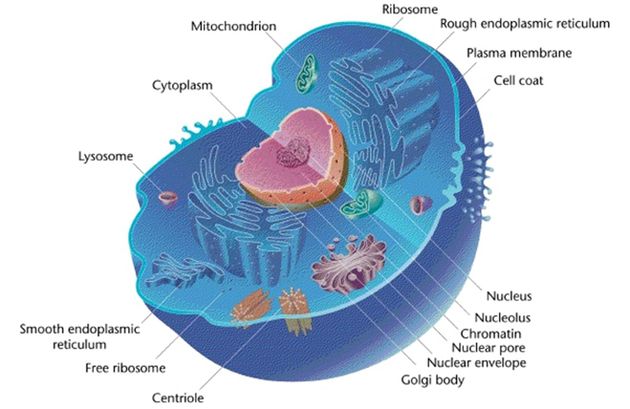
He explains each organelle's function including the nucleus, nucleolus, nuclear envelope, nuclear pore, chromatin, dna, cytoskeleton, lysosome, perixosome, rough and smooth endoplasmic reticulum, golgi apparatus, ribsomes, vesicles. The cytoskeleton organizes other constituents of the cell, maintains the cell's shape, and is responsible for the locomotion of the cell itself and the movement of the various organelles within it. The cytoskeleton of a biological cell is the framework of tiny tubes and filaments that forms the internal structure of the see the diagram to see how these are arranged. Unlike the eukaryotic cells of plants and fungi, animal cells do not have a cell wall. It is a network of protein fibers supporting cell shape and anchoring organelles within the cell. They are stained with fluorescent labels to help visualise the cytoskeleton with microtubules (green), actin filaments (red), and the nucleus (blue). In addition to providing structural support, it's also involved in different types of movements (where it anchors various cellular structures like the flagellum) as well as the movement of cellular substances. A cytoskeleton is present in the cytoplasm of all cells, including bacteria, and archaea. An animal cell ranges in size from 10 to 30 µm. Protrusions (cilia and flagella) in some cells. This provides a cellular scaffolding that arranges the cellular organization into. Golgi apparatus cytoskeleton smooth endoplasmic reticulum nucleolus nuclear envelope nucleus 4 cytokinesis mitosis cell cycle s g1g1 g2g2 cell cycle. It forms cellular appendage, for example;
5 metaphase anaphase telophase kinetochore microtubule early metaphase metaphase. In the space below, record the venn diagram you created in the simulation comparing a typical animal cell and plant cell. In animal cells the mtoc is called the centrosome. It is a complex, dynamic network of interlinking protein filaments that extends from the cell nucleus to the cell membrane.1 the cytoskeletal systems of different organisms are composed of. Contains microfilaments, microtubules, intermediate filaments.
The Eukaryotic Cell Cytoskeleton - ScienceAid from scienceaid.net They are stained with fluorescent labels to help visualise the cytoskeleton with microtubules (green), actin filaments (red), and the nucleus (blue). It is a network of protein fibers supporting cell shape and anchoring organelles within the cell. An animal cell ranges in size from 10 to 30 µm. It forms cellular appendage, for example; The cytoskeleton is made up of two words cyto and skeleton. .the prefix cyto means cell so cytoskeleton simply means the skeleton of the cell and although pretty much all cells on the cytoskeleton that's found in animal cells so the cytoskeleton. The diagram, like the one above, will include labels of the major parts of an animal cell including the cell membrane, nucleus, ribosomes, mitochondria, vesicles, and cytosol. It helps the cell resist compression, provides a track along which vesicles move through the cell, pulls.
Printable animal cell diagram to help you learn the organelles in an animal cell in preparation for your test or quiz.
Microtubules, microfilaments and intermediate filaments. The diagram, like the one above, will include labels of the major parts of an animal cell including the cell membrane, nucleus, ribosomes, mitochondria, vesicles, and cytosol. In animal cells the mtoc is called the centrosome. Several cellular structures are built around a core of cytoskeletal proteins. The different types of the cytoskeleton are actin filaments, intermediate filaments. A cell's cytoskeleton ensures stability, energy, and motility. The cytoskeleton organizes other constituents of the cell, maintains the cell's shape, and is responsible for the locomotion of the cell itself and the movement of the various organelles within it. Microfilaments are the thinnest of all the cytoskeletal. Internal protein framework that provides support and structure for the cell; He explains each organelle's function including the nucleus, nucleolus, nuclear envelope, nuclear pore, chromatin, dna, cytoskeleton, lysosome, perixosome, rough and smooth endoplasmic reticulum, golgi apparatus, ribsomes, vesicles. Summary of the structure and function of a eukaryotic cell. It is a complex, dynamic network of interlinking protein filaments that extends from the cell nucleus to the cell membrane.1 the cytoskeletal systems of different organisms are composed of. An animal cell diagram is a great way to learn and understand the many functions of an animal cell.
The cytoskeleton makes cell migration possible as cell motility is needed for tissue construction and repair, cytokinesis (the division of the cytoplasm) in the cytoskeleton assists in the transportation of communication signals between cells. Diagram of animal cell, created with biorender.com. All cells have a cytoskeleton. This provides a cellular scaffolding that arranges the cellular organization into. Contains microfilaments, microtubules, intermediate filaments.
cell diagrams - biology 11 portfolio from vanessawhitebio11.weebly.com Animal cell to cruise ship analogy > . The result is two centrosomes microtubules (and centrioles) are part of the cytoskeleton. In animal cells, two networks of intermediate filaments provide the nucleus with mechanical support: In cell biology, the cytoskeleton is a system of fibrillar structures that pervades the cytoplasm. In the complete animal cell centrosome, the two centrioles are arranged such that one is perpendicular to the other. Summary of the structure and function of a eukaryotic cell. Golgi apparatus cytoskeleton smooth endoplasmic reticulum nucleolus nuclear envelope nucleus 4 cytokinesis mitosis cell cycle s g1g1 g2g2 cell cycle. It is a complex, dynamic network of interlinking protein filaments that extends from the cell nucleus to the cell membrane.1 the cytoskeletal systems of different organisms are composed of.
Cytoskeleton structure consists of framework of filaments and tubules to help in function of cytoskeleton i.e., provide support and shape to the cell.
An animal cell diagram is a great way to learn and understand the many functions of an animal cell. The cytoskeleton is made up of two words cyto and skeleton. In animal cells the mtoc is called the centrosome. In the complete animal cell centrosome, the two centrioles are arranged such that one is perpendicular to the other. This image shows some animal cells. Microtubules, microfilaments and intermediate filaments. The cytoskeleton organizes other constituents of the cell, maintains the cell's shape, and is responsible for the locomotion of the cell itself and the movement of the various organelles within it. An animal cell ranges in size from 10 to 30 µm. Cytoskeleton, a system of filaments or fibers that is present in the cytoplasm of eukaryotic cells. A cytoskeleton is present in the cytoplasm of all cells, including bacteria, and archaea. Printable animal cell diagram to help you learn the organelles in an animal cell in preparation for your test or quiz. Golgi apparatus cytoskeleton smooth endoplasmic reticulum nucleolus nuclear envelope nucleus 4 cytokinesis mitosis cell cycle s g1g1 g2g2 cell cycle. In cell biology, the cytoskeleton is a system of fibrillar structures that pervades the cytoplasm.
Share :
Post a Comment
for "Animal Cell Diagram Cytoskeleton / Animal Cell Diagram - Labeled - Tim van de Vall / Unlike the eukaryotic cells of plants and fungi, animal cells do not have a cell wall."


Post a Comment for "Animal Cell Diagram Cytoskeleton / Animal Cell Diagram - Labeled - Tim van de Vall / Unlike the eukaryotic cells of plants and fungi, animal cells do not have a cell wall."